The University of Sydney Takes a Leap Towards Functional 3D Printed Human Tissues
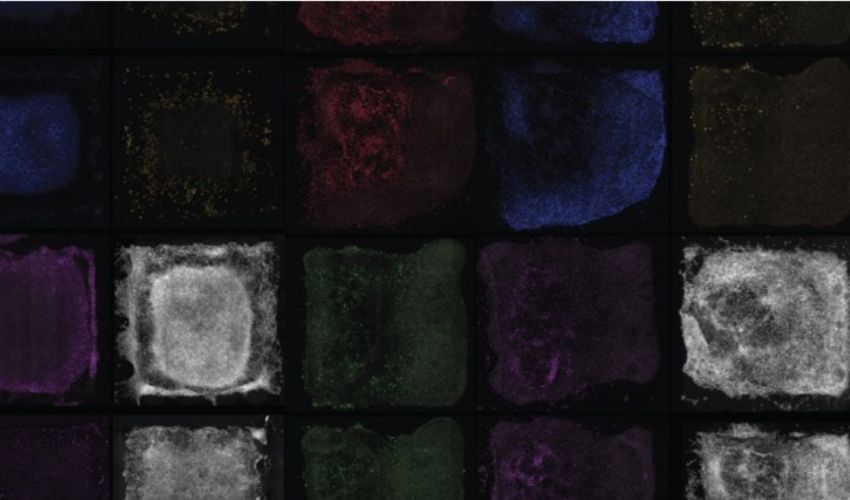
A recent breakthrough, which seems to have been lifted straight from science fiction books, has moved us closer to 3D printing human functional tissues. A collaborative effort between bioengineers and biomedical scientists from the University of Sydney and the Children’s Medical Research Institute (CMRI) at Westmead has harnessed the power of 3D photolithographic printing to unveil a sophisticated method that ushers in a new era of tissue engineering. This method involves creating a precisely crafted environment to mirror the architectural details found in human organs.
Under the guiding expertise of Professor Hala Zreiqat and Dr. Peter Newman from the University of Sydney’s School of Biomedical Engineering, and with developmental biologist Professor Patrick Tam at the helm of the CMRI’s Embryology Research Unit, this pioneering technique orchestrates a remarkable transformation. The versatile building blocks of the life are directed to metamorphose and become specialized cells. These cells then harmoniously assemble themselves into a structure which closely resembles an organ.
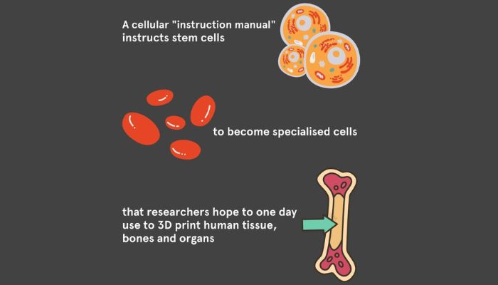
A cellular “guidebook” directs stem cells to transform into specialized cells with the future goal of 3D printing human tissue, bones, and organs (Photo credits: The University of Sydney)
The cells’ intricate microenvironment is choreographed in a similar way to how a needle on a record player navigates the grooves to create music. This complex ballet is orchestrated by strategically placed proteins and precise mechanical signals, which effectively replicates the complex developmental processes within the body.
The culmination of this endeavor is a new paradigm that has been aptly described as an “instruction manual” for cells. Professor Hala Zreiqat describes the transformative nature of this technique by declaring: “Our new method serves as an instruction manual for cells, allowing them to create tissues that are better organized and more closely resemble their natural counterparts. This is an important step towards being able to 3D print working tissue and organs.” This “instruction manual” transcends mere biological guidance, ushering in an era where human ingenuity meets the complexity of life itself.
Imagine for a minute the art of building a structure from a variety of different components. Dr. Peter Newman uses a similar analogy, asking us imagine building a Lego Castle by randomly scattering the blocks on a desk. A castle would be more likely to look like a mess of blocks if there was no plan. In the same way, creating functional tissues out of cells requires a detailed roadmap. The “instruction manual” provides this guidance, ensuring that cells harmoniously organize themselves to create tissues that closely mimic their natural counterparts.

Dr. Newman explained the need for precise guidance when creating tissues from a cell, similar to assembling a building from different components. (Photo credit Pixabay/Aldarami).
This breakthrough in tissue engineering has a multitude of implications. The understanding of the complex tissue structures called organoids and their development and functionality not only helps to unravel the mysteries behind disease origins, but also pushes the boundaries of cell and gene therapies. This breakthrough has the potential to transform lives and revolutionize the treatment of conditions such as macular degeneration or inherited disorders which lead to loss of retinal cells. The future involves the advancement of regenerative medicines, discovering novel approaches to treat a range diseases, and inspiring hope in those who imagine a better, healthier tomorrow. Click HERE for more information about the study.
What do you think of this new method for 3D printing human tissues? Let us know in a comment below or on our LinkedIn, Facebook, and Twitter pages! Don’t forget to sign up for our free weekly Newsletter here, the latest 3D printing news straight to your inbox! You can also find all our videos on our YouTube channel.
*Cover photo credit: The University of Sydney